NUHS Institutions will NEVER ask you to transfer money or disclose bank details over a call. If in doubt, call the 24/7 ScamShield helpline at 1799, or visit the ScamShield website at www.scamshield.gov.sg.
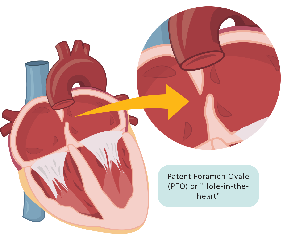
Patent Foramen Ovale (PFO) is a congenital heart condition commonly known as a “hole in the heart”. The “hole” is a small, flaplike opening between the upper chambers of the heart called the atria that is required only when one is a foetus. This hole usually closes after birth when the foetus’ lungs begin to work.
When the hole does not close as it normally should after birth, it can sometimes cause small blood clots to pass between the heart chambers, potentially obstructing blood flow in vessels and causing strokes when they reach the brain.
What are the symptoms?
PFO occurs in about 1 in 4 people. It usually has no symptoms if there is no blockage of blood vessels by blood clots that pass through the “hole”. PFO is often discovered during tests for other health problems.
Possible risks of PFO when a blockage occurs include:
Stroke
Transient Ischemic Attack, also known as a mini-stroke, that impairs one’s ability to move limbs and also affects vital organs
Heart Attack (Myocardial Infarction)
Low blood oxygen levels
Who is at a higher risk of PFO?
PFO is frequently found in: -Divers with decompression illness (air embolism) -People who have had a stroke of unknown cause (cryptogenic stroke)
Despite this, anyone can have PFO, regardless of gender, ethnicity, race, or lifestyle, as PFO is a congenital cardiac condition that arises from birth.
How is it diagnosed?
Your healthcare provider will first ask about your medical conditions and do a physical examination. If your healthcare provider suspects PFO, they may recommend conducting additional diagnostic tests.
What are the tests?
Transthoracic echocardiogram (TTE):
What is it?
The most common type of echocardiogram that uses ultrasound to produce images of your heart. This test is non-invasive as no part of the instrument is inserted into the body.
Why is it done?
To view the structure and size of your heart’s chambers and assess for abnormalities.
Does it hurt?
This test is painless.
Potential risks and complications?
TTE is extremely safe. No clinically important adverse effects have been reported, even with repeated examinations. Pregnant women may also undergo TTE, without additional risk to themselves or the unborn child.
Transoesophageal echocardiogram (TEE):
What is it?
TEE is a type of echocardiogram that capture images of your heart via ultrasounds waves and the use of a thin, flexible tube down your oesophagus. Unlike other types of echo tests, this test creates pictures from inside your body.
Why is it done?
TEE is usually done after an echocardiogram performed from the outside of your chest (TTE), to obtain an even more detailed visualisation of your heart’s structures to assess for abnormalities.
Does it hurt?
Local anaesthetic will be applied to your throat to reduce discomfort during the test.
Potential risks and complications?
This test has minimal risks; some may experience slight discomfort or a sore throat after the test.
Transcranial Doppler (TCD) ultrasound:
What is it?
A non-invasive test that uses ultrasound to identify issues impacting blood circulation to and within the brain.
Why is it done?
To detect possible stroke caused by blood clots, narrowed blood vessels, haemorrhage and more.
Does it hurt?
No, TCD Ultrasound is typically painless.
Potential risks and complications?
There are usually no significant risks or complications associated with this test as it does not utilise radiation, as X-ray tests do. It is considered a quick and safe diagnostic tool.
What are the treatments?
Most PFO patients do not require treatment unless they are at risk of stroke or blood clots. Treatments include medications and closure of the hole with a catheter or through surgery.
Medications:
What is it?
Prescribed to reduce blood clots in the body.
What to expect?
With a reduction of blood clots in blood vessels, risks of blood clot related critical conditions such as stroke or pulmonary embolism is decreased.
What to prepare?
Please follow the prescribed dosage and schedule as advised by your doctor.
Success rates:
This would vary based on one’s compliance to medication-taking and the severity of the condition.
Potential risks & complications:
Side effects vary; kindly consult with your doctor.
When to call the doctor:
If there are any unexpected side effects or if symptoms persist.
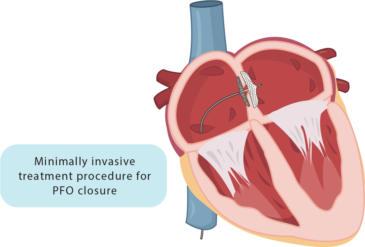
PFO Closure through Minimally Invasive Transcatheter Repair:
What is it?
Closing of the hole between the heart’s two upper chambers through the plugging in of a permanent device.
What to expect?
A thin, flexible, and long hollow tube called the catheter will be inserted into the large vein in the groin and advanced through the blood vessel to access the heart. After which, a closure device will be used to seal the hole. Upon closing the hole with the PFO closure device, the body’s tissue will begin to grow over the device. In three to six months, the heart tissues will eventually fully cover the device to form part of the heart’s wall.
What to prepare?
Pre-procedure evaluation will be required, including undergoing several diagnostic tests such as: -Electrocardiogram (ECG) -Echocardiogram -Chest X-ray -Blood tests
Medications would also have to be taken to prevent blood clots before the PFO closure procedure.
Potential risks & complications:
Applicable procedure risks may include: •Blood clotting at procedure site •Blood clot in the lungs (Pulmonary Embolism) •Blood vessel damage •Movement of the closure device •Bacterial infection in the heart (Endocarditis) •PFO device may fail to close •Atrial Fibrillation •Arrhythmia
When to call the doctor:
Contact your healthcare provider if there are signs of infection at the puncture site, severe pain, or other post-procedure complications.
Living with PFO
You can keep your heart healthy and reduce your risk of stroke by: -Avoid alcohol or use of recreational drugs -Eat a heart-healthy diet -Keep your blood pressure and blood cholesterol under control -Maintain a healthy weight -Quit smoking and the use of tobacco products -Reduce your risk of blood clots by having an active lifestyle