Heart Information

Enlarged Heart (Cardiomegaly)

2026/01/16
What is Enlarged Heart (Cardiomegaly)?
Cardiomegaly, or enlarged heart, refers to a heart that is bigger in size than normal. It is not a disease itself, but a sign of an underlying condition that causes the heart muscle to thicken or stretch. It may affect the whole heart or just one part and can lead to decreased pumping efficiency or heart failure if not managed.

Cardiomegaly can be temporary or permanent, depending on its underlying cause. It often occurs when the heart must exert more effort to pump blood, similar to how muscles grow with exercise.
What are the symptoms?

Some people may have no symptoms. When symptoms occur, they may include:

  • Shortness of breath, especially when lying flat or during physical activity
  • Swelling in the legs, ankles, or abdomen
  • Fatigue
  • Irregular or rapid heartbeat (palpitations)
  • Dizziness or fainting
  • Weight gain from fluid retention
  • Chest pain (less common)
How do we evaluate it?

Doctors diagnose an enlarged heart based on:

  • A physical examination
  • Medical history review
  • Imaging tests to assess the size and function of the heart
  • Tests to uncover the underlying cause (e.g., high blood pressure, valve disease, cardiomyopathy)
What are the tests to diagnose Cardiomegaly?

1. Blood Tests

What is it?
Blood samples are taken to check for signs of heart failure, thyroid disorders, anaemia, or infection.

Why is it done?
To find the cause or consequences of the enlarged heart.

Does it hurt?
Only a brief pinch from the needle is required to draw blood for the laboratory test.

Potential risks and complications?
Minor bruising or discomfort at the needle site.

2. Electrocardiogram (ECG or EKG)

What is it?
An electrocardiogram is a simple, non-invasive test that records the heart's electrical activity and rhythm.

Why is it done?
To detect irregular heart rhythms and identify arrhythmias.

Does it hurt?
No, an ECG is painless and involves the placement of electrodes on the skin's surface to detect electrical signals. 

Potential risks or complications?
There are virtually no risks or complications associated with ECGs. It's a quick and safe diagnostic tool.

3. Chest X-ray

What is it?
A basic imaging test that captures the size and shape of the heart, lungs, blood vessels, airways and bones of the chest. Chest X-rays can also reveal fluid in or around your heart or lungs or air surrounding a lung. 

Why is it done?
Often, the first sign of the enlarged heart is picked up through an X-ray. A chest X-ray can also be used to check how you are responding to treatment. 

Does it hurt?
X-rays are generally painless as no sensation can be felt as the radiation passes through the body.

Potential risks and complications?
One may be concerned about radiation exposure from chest X-rays, especially if there is a need to take them regularly. However, the amount of radiation from a chest X-ray is even lower than natural sources of radiation found in the environment where one is generally exposed to. The benefits of an X-ray also outweigh the risks, and a protective apron will be provided during the test if multiple images are required. Kindly inform the staff if you are pregnant and further measures will be done to protect the belly area from radiation.

4. Echocardiogram (ECHO)

What is it?
An echocardiogram is a non-invasive imaging test that uses high-frequency sound waves to create detailed images of the heart's structure and function. 

Why is it done?
Echocardiograms are performed to access the size of the heart and evaluate its impact on heart function. 

Does it hurt?
No, an echocardiogram is a painless procedure that involves the application of a gel on the chest and the use of a handheld transducer to capture images. It's generally well-tolerated by patients, including children.

Potential risks and complications?
There are no significant risks or complications associated with echocardiograms. It is a safe and widely used diagnostic tool for cardiac conditions.

5. CT Scan

What test is it?
A CT scan first uses X-rays to produce detailed images of the body and after which, a computer is used to create cross-sectional images (known as slices) of the bones, tissues, and blood vessels. Images from a CT scan can show more details than that of a regular X-ray.

Why is it done?
CT scans provide more detailed images of the heart, especially that of the blood vessels of the heart (coronary arteries).

Does it hurt?
The test itself is painless, but some people may be sensitive to the contrast dye used in selected CT scans, which can cause mild discomfort.

Potential risks and complications?
The risks can include allergic reactions to the contrast dye and exposure to ionizing radiation. However, the benefits usually outweigh the risks in most cases.

6. Cardiovascular MRI

What test is it?
MRI is a non-invasive diagnostic approach that uses a magnetic field and computer-generated radio waves to create detailed images of organs, bones, and soft tissues in the body.

Why is it done?
To provide detailed images of the heart, including tissue that cannot be seen on a conventional x-ray, to help in diagnosing the underlying cause of cardiomegaly. 

Does it hurt?
The scan itself is painless as the patient does not feel the magnetic field and there are no moving parts around the patient. However, some individuals may experience possible feelings of claustrophobia in the machine or be sensitive to the sounds produced by the magnets during the scan, which can cause mild discomfort. Wearing earplugs may help to block the noise. 

Potential risks & complications?
MRI is considered safe for most people as it does not use radiation. However, patients with certain medical devices or conditions may not be eligible for an MRI as MRI uses a strong magnetic field – the presence of metal objects in the body can be a safety hazard or distort the MRI images.

7. Cardiac Catheterisation and Angiography

What is it?
Angiography is a diagnostic test that involves injecting a contrast dye into the bloodstream and using X-rays to create real-time images of the selected blood vessel.

Why is it done?
Angiography is used to provide precise imaging and information about a vessel’s anatomy, as blood vessel blockage is a common cause of a weak and enlarged heart. The injected contrast material makes it easy to see where blood is moving and where blood vessel blockages or damages are. During the procedure, pressures in the heart can be measured as well.

Does it hurt?
The procedure may cause some discomfort, especially when the contrast dye is injected. Local anaesthesia will be administered for improved comfort.

Potential risks & complications?
There are risks associated with angiography, including allergic reactions to the contrast dye, bleeding at the catheter insertion site, and a small risk of damage to blood vessels. The procedure is considered safe but may carry more risks compared to non-invasive imaging method.

What are the treatments?

Treatment focuses on managing the underlying cause of the enlarged heart and preventing complications.

1. Medications

You will be prescribed a combination of drugs such as:

  • ARN-I/ ACE inhibitors/ ARBs: lower blood pressure and reduce strain on the heart 
  • Beta-blockers: slow the heart rate and improve heart function
  • Aldosterone antagonists: reduce fluid retention and improve heart function
  • SGLT-2 inhibitors: promote sugar excretion, improving heart and kidney outcomes
  • Diuretics: help remove excess fluid in the body

Home care advice: Most people take multiple medications daily. Do note to take your medications on time and track any side effects. Please contact your care team if you experience new side effects, worsening swelling, chest pain or irregular heartbeat.

2. Treating Underlying Conditions

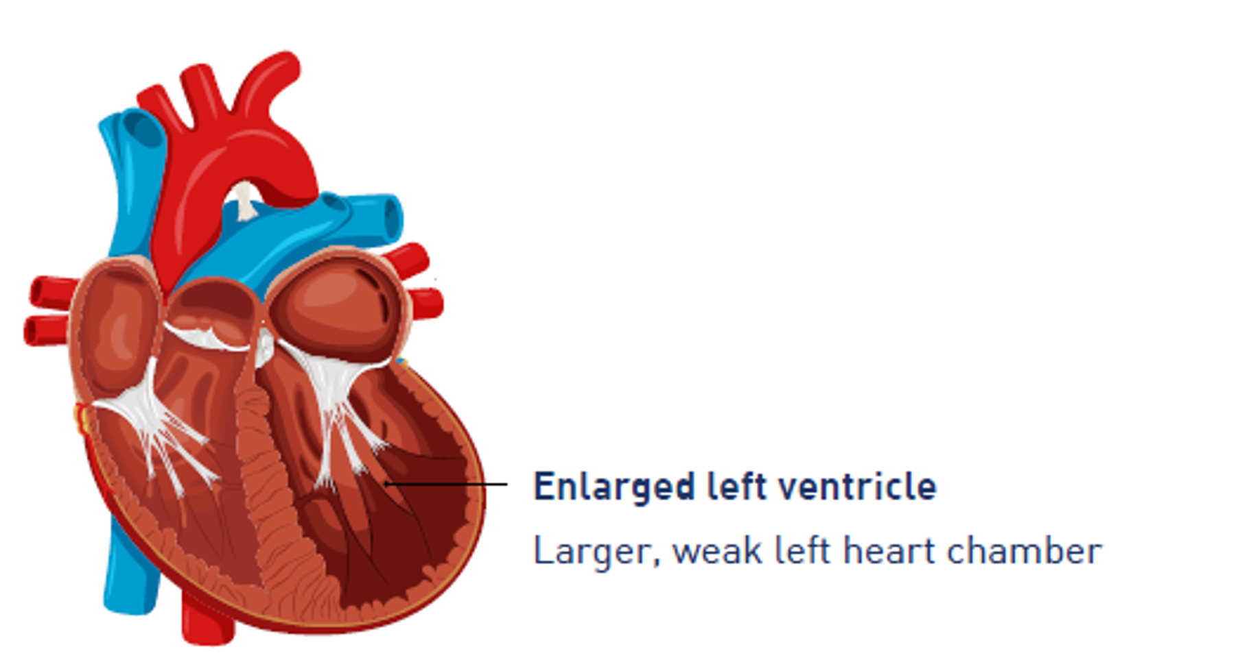
Cardiomegaly, or an enlarged heart, can be caused by several underlying issues, including:

  • High blood pressure
  • Valve disease
  • Thyroid conditions
  • Anaemia

If these root causes are effectively managed, improvements in both heart size and related symptoms can be expected. To support heart health, patients should focus on making lifestyle changes and adhering to prescribed medications. Should there be any signs of worsening underlying conditions, please contact your care team immediately. 

3. Lifestyle Changes

You are strongly recommended to have a low-sodium diet, restrict your fluid intake, quit smoking, limit alcohol consumption, lose weight, and engage in regular light exercise as part of your everyday lifestyle.

Through such lifestyle changes, not only will your medical symptoms reduce over time, quality of life will also be improved.

Home care advice: Please monitor your weight, avoid salty foods, and track your symptoms. If you feel increasingly breathless, or experience swelling of the body, please contact your care team immediately.

4. Surgery or Procedures (if needed)

Cardiac surgery is a treatment option depending on the underlying cause of cardiomegaly, particularly those experiencing structural heart problems or heart failure. This may involve valve repair or replacement surgeries, Coronary Artery Bypass Grafting (CABG), or the implantation of devices such as pacemakers or defibrillators. 

Patients can expect to undergo pre-operative tests and will typically require a hospital stay, followed by a recovery period. As with any surgery, there are risks involved, including bleeding, infection, and potential device failure. After the operation, home care is essential, which includes monitoring the surgical site and adhering to activity restrictions. It's important to contact your care team if there are signs of fever, wound complications, shortness of breath, or heart palpitations during recovery.

Last updated on
Best viewed with Chrome 79.0, Edge 112.0, Firefox 61.0, Safari 11
National University Health System
  • National University Hospital
  • Ng Teng Fong General Hospital
  • Alexandra Hospital
  • Tengah General and Community Hospital
  • Jurong Community Hospital
  • National University Polyclinics
  • Jurong Medical Centre
  • National University Cancer Institute, Singapore
  • National University Heart Centre, Singapore
  • National University Centre for Oral Health, Singapore
  • NUHS Diagnostics
  • NUHS Pharmacy
  • NUHS Regional Health System Office
  • NUS Yong Loo Lin School of Medicine
  • NUS Faculty of Dentistry
  • NUS Saw Swee Hock School of Public Health
Back to Top