Diagnosis involves a combination of medical history evaluation, physical examination, and diagnostic tests to assess heart function and structure. Key diagnostic approaches include:
1. Medical History and Physical Exam:
Assessing risk factors such as long-standing hypertension, lifestyle habits, and family history.
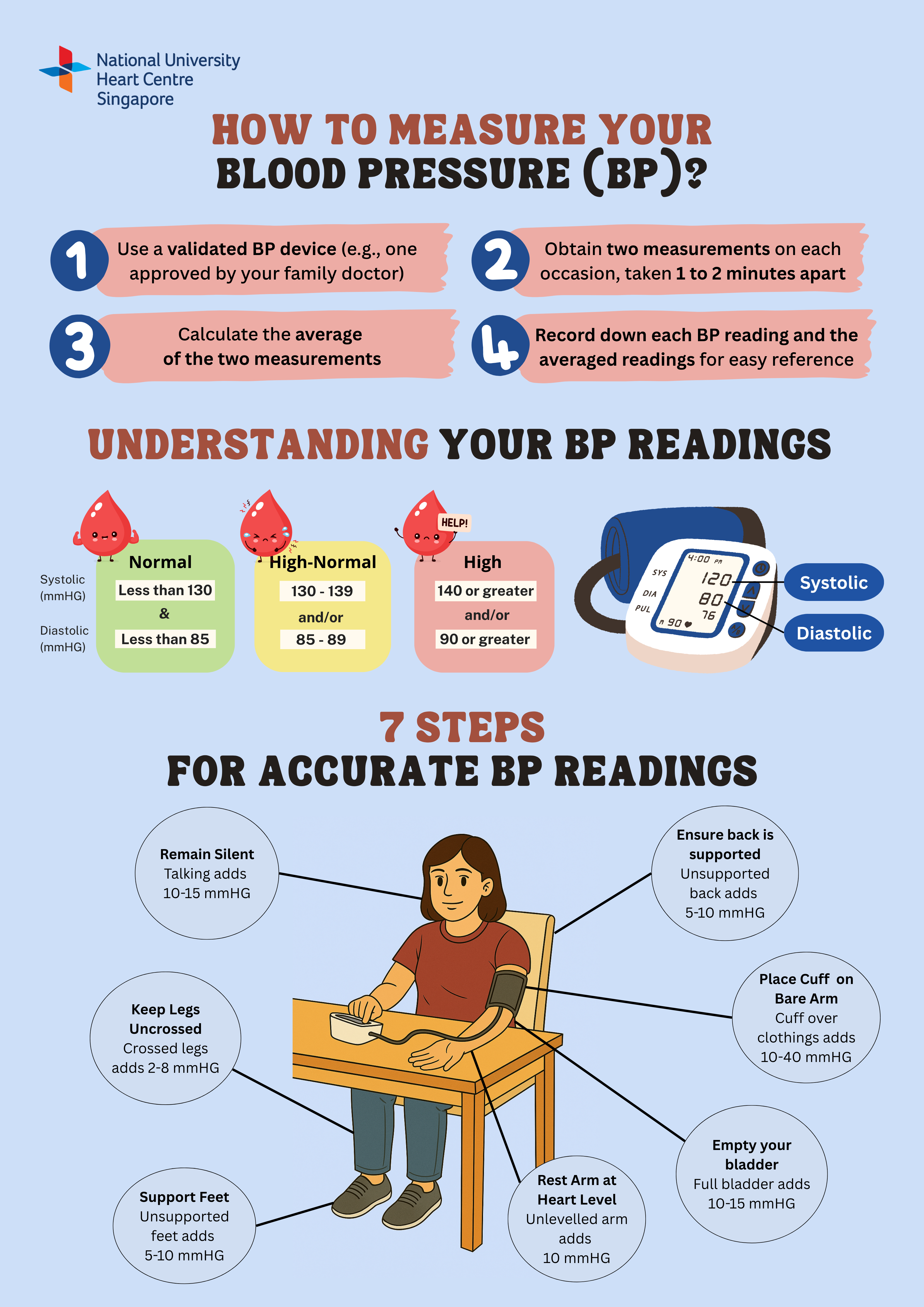
2. Blood Pressure Measurement:
Consistent readings above 140/90 mmHg when measured on two separate occasions indicate hypertension.
3. Blood Tests:
Used to assess kidney function, cholesterol levels, and markers of heart strain including Natriuretic Peptide (NT-proBNP).
4. Electrocardiogram (ECG):
What is it?
An electrocardiogram is a simple, non-invasive test that records the heart's electrical activity and rhythm.
Why is it done?
To detect irregular heart rhythms and structural heart diseases.
Does it hurt?
No, an ECG is painless and involves the placement of electrodes on the skin's surface to detect electrical signals.
Potential risks or complications?
There are virtually no risks or complications associated with ECGs. It's a quick and safe diagnostic tool.
5. Blood Tests:
What is it?
A basic imaging test that captures the size and shape of the heart, lungs, blood vessels, airways and bones of the chest. Chest X-rays can also reveal fluid in or around your heart or lungs or air surrounding a lung.
Why is it done?
The image helps determine whether you have heart problems, a collapsed lung, pneumonia, emphysema, cancer or any of several other conditions. For example, changes in the size and shape of your heart may indicate heart failure, fluid around the heart or heart valve problems. A chest X-ray can also be used to check how you are responding to treatment.
Does it hurt?
X-rays are generally painless as no sensation can be felt as the radiation passes through the body.
Risks and complications?
One may be concerned about radiation exposure from chest X-rays, especially if there is a need to take them regularly. However, the amount of radiation from a chest X-ray is even lower than natural sources of radiation found in the environment where one is generally exposed to. The benefits of an X-ray also outweigh the risks, and a protective apron will be provided during the test if multiple images are required. Kindly inform the staff if you are pregnant and further measures will be done to protect the belly area from radiation.
6. Echocardiogram
What is it?
An echocardiogram is a non-invasive imaging test that uses high-frequency sound waves to create detailed images of the heart's structure and function.
Why is it done?
Echocardiograms are performed to confirm the size of the heart and evaluate its impact on heart function.
Does it hurt?
No, an echocardiogram is a painless procedure that involves the application of a gel on the chest and the use of a handheld transducer to capture images. It's generally well-tolerated by patients, including children.
Potential risks and complications?
There are no significant risks or complications associated with echocardiograms. It is a safe and widely used diagnostic tool.
7. Cardiovascular Mass Resonance Imaging (MRI)
What test is it?
MRI is a non-invasive diagnostic approach that uses a magnetic field and computer-generated radio waves to create detailed images of organs, bones, and soft tissues in the body.
Why is it done?
To provide detailed images of the heart, including tissue that cannot be seen on a conventional x-ray, to help in diagnosing the underlying cause of heart failure, particularly heart failure secondary to diseases of the heart muscles.
Does it hurt?
The scan itself is painless as the patient does not feel the magnetic field and there are no moving parts around the patient. However, some individuals may experience possible feelings of claustrophobia in the machine or be sensitive to the sounds produced by the magnets during the scan, which can cause mild discomfort. Wearing earplugs may help to block the noise.
Potential risks & complications?
MRI is considered safe for most people as it does not use radiation. However, patients with certain medical devices or conditions may not be eligible for an MRI as MRI uses a strong magnetic field – the presence of metal objects in the body can be a safety hazard or distort the MRI images.