Diagnosis involves a combination of clinical evaluation, laboratory tests, imaging studies, and tissue biopsy:
1. Medical history and physical examination of the body to assess symptoms and risk factors
2. Blood and Urine Tests
What is it?
Laboratory analyses are conducted to detect abnormal light-chain proteins (free light-chain assay) and assess organ function.
Why is it done?
To identify the presence of monoclonal light-chain proteins that are indicative of AL Amyloidosis.
Does it hurt?
Blood draw involves minor discomfort; urine collection is non-invasive.
Potential risks and complications:
There are minimal risks and complications associated with blood and urine tests, except the possibility of slight bruising at the blood draw site.
3. Electrocardiogram (ECG)
What is it?
An electrocardiogram is a simple, non-invasive test that records the heart's electrical activity and rhythm.
Why is it done?
To detect irregular heart rhythms and screen for structural heart disease.
Does it hurt?
No, an ECG is painless and involves the placement of electrodes on the skin's surface to detect electrical signals.
Potential risks or complications?
There are virtually no risks or complications associated with ECGs. It's a quick and safe diagnostic tool.
4. Echocardiogram
What is it?
An echocardiogram is a non-invasive imaging test that uses high-frequency sound waves to create detailed images of the heart's structure and function.
Why is it done?
Echocardiograms are performed to confirm the size of the heart and evaluate its impact on heart function. It is a key tool for us to evaluate for heart involvement in light chain amyloidosis.
Does it hurt?
No, an echocardiogram is a painless procedure that involves the application of a gel on the chest and the use of a handheld transducer to capture images. It's generally well-tolerated by patients, including children.
Potential risks and complications?
There are no significant risks or complications associated with echocardiograms. It is a safe and widely used diagnostic tool.
5. Cardiovascular MRI
What test is it?
MRI is a non-invasive diagnostic approach that uses a magnetic field and computer-generated radio waves to create detailed images of organs, bones, and soft tissues in the body.
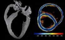
Why is it done?
To provide detailed images of the heart, including tissue that cannot be well evaluated on a conventional x-ray or echocardiogram. This will help in the diagnosis of cardiac amyloidosis.
Does it hurt?
The scan itself is painless as the patient does not feel the magnetic field and there are no moving parts around the patient. However, some individuals may experience possible feelings of claustrophobia in the machine or be sensitive to the sounds produced by the magnets during the scan, which can cause mild discomfort. Wearing earplugs may help to block the noise.
Potential risks & complications?
MRI is considered safe for most people as it does not use radiation. However, patients with certain medical devices or conditions may not be eligible for an MRI as MRI uses a strong magnetic field – the presence of metal objects in the body can be a safety hazard or distort the MRI images.
6. Heart Muscle Biopsy
What is it?
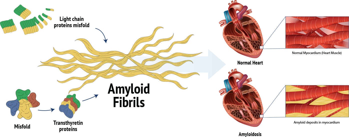
A medical procedure that involves taking a small sample of tissue from the heart for examination under a microscope. It is commonly used to diagnose heart muscle diseases, including cardiac amyloidosis, which is characterized by the abnormal accumulation of amyloid proteins in tissues and organs, in particular the heart.
Why is it done?
To confirm the presence of amyloid deposits in the heart, which helps in diagnosing amyloidosis. It can also help determine the type of amyloidosis, which is crucial for deciding the appropriate treatment.
Does it hurt?
This is an invasive procedure. Local anaesthesia is used to numb the site of biopsy catheter entry, so patients may feel some pressure or a brief sting but should not experience significant pain.
Potential risks and complications:
While cardiac biopsies are generally safe, there are some potential risks and complications, including bleeding at the biopsy site, infection, pain or discomfort in the area where the biopsy was taken, or damage to surrounding tissues or heart. Overall, the benefits of obtaining a definitive diagnosis often outweigh the risks associated with the procedure.