Learning Abroad, Caring At Home
How insights from a cardiac imaging fellowship are transforming patient care at NUHCS
PULSE Issue 47 | July 2026
The National University Heart Centre, Singapore (NUHCS) is committed to providing world-class cardiac care. A key part of this mission is fostering growth of our cardiologists by offering global exchange opportunities, also known as fellowships.
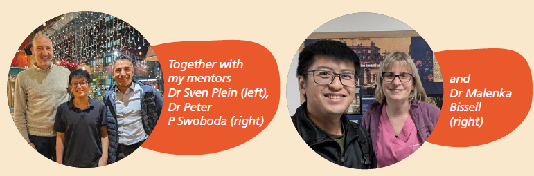
PULSE speaks to Dr Sia Ching Hui, Consultant, Department of Cardiology, NUHCS, who recently completed a fellowship in cardiac Magnetic Resonance Imaging (MRI)1 at Leeds General Infirmary in the United Kingdom (UK). What began as an opportunity to deepen his expertise in advanced cardiac imaging became a journey that changed the way he thinks about patient care.
What is a Fellowship and why do public healthcare institutes offer this?
A fellowship is an advanced training programme designed for specialists to delve deeper into their area of expertise, allowing them to refine skills, master cutting-edge techniques, and gain the knowledge and confidence to manage the most complex cases within their specialty. Many public healthcare institutes, like NUHCS, offer doctors with fellowship opportunities for patients to access top-tier care from highly skilled experts, and foster groundbreaking research with fresh ideas.
As a cardiologist, I have always been fascinated by what we can learn about the heart through imaging. When I was given the opportunity to further develop my expertise, I expected to learn how to interpret more complex scans. What I did not expect was how the experience would reshape the way I think about patient care. It changed not only how I read the scanned images, but also how I understand the patient’s journey and the decisions that follow. More importantly, it reminded me that behind every scan is a person waiting for answers, and every report we write has the potential to influence the course of someone’s life.
Seeing What Matters
One of the most valuable lessons I learned was how to write better reports. Every scan I reviewed was discussed with a senior specialist, word by word. In the beginning, my reports were long and descriptive, recounting everything I saw. Over time, I was challenged to think differently: what is the one message the clinician needs? That shift changed everything. Reports became clearer, more concise, and more purposeful. I have since brought this mindset back to NUHCS, where we are working towards reports that help clinicians make decisions quickly rather than hunting for meaning.
The fellowship also offered me a close look at emerging imaging technologies. One example was 4D Flow MRI2, which visualises blood movement through the heart in three dimensions over time. Rather than inferring how the blood flows, we can observe it directly as it moves through a narrowed valve or an abnormal vessel. For patients with valve disease3 or congenital heart conditions4, this provides valuable insights into why their condition may be worsening and when intervention is needed. Seeing it used routinely highlighted its potential for patients in Singapore. We are now taking steps to introduce this technology at NUHCS.
Another important development was quantitative perfusion imaging5, which measures blood flow to the heart muscle. This goes beyond visual assessment and is particularly helpful for patients who experience chest pain despite having no obvious blockages in their arteries. Many of these patients have previously been told that their tests were normal, leaving them without answers. By identifying reduced blood flow in the small vessels, we can provide clearer explanations and guide treatments more effectively.
Did you know that a scan result can change someone’s life?
It may determine whether a patient needs surgery, whether a young athlete can safely return to sport, or whether a family should be screened for an inherited condition. With this comes a responsibility to ensure every report is clear, accurate and meaningful.
Imaging with a Human Touch
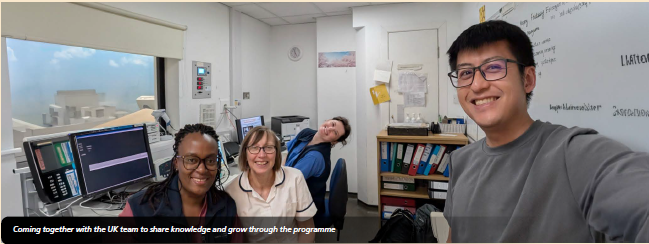
What stood out most during the fellowship was not the technology, but the emphasis on patient experience. Children were scanned using breathing techniques that allowed them to breath normally, avoiding repeated breath-holding. They could watch cartoons during the scan, and parents were encouraged to stay close by. Adults lay in softly lit rooms and watched Netflix while their heart scans were being performed. Behind the scenes, radiographers carried out complex stress scans, while physicists worked alongside doctors to improve efficiency without compromising on image quality. These details made a meaningful difference. Children required less sedation6. Adults felt calmer and were more cooperative. Scans were completed more efficiently, and image quality improved. It showed me that advanced technology and compassionate care can go hand-in-hand.
Bringing These Lessons Home
Back at NUHCS, my focus has been on adapting these lessons to our local setting. While our workflows and patient needs may differ from the UK, the principles remain relevant. We are working to:
- Streamline imaging protocols so patients spend less time in the scanner
- Introduce structured co-reporting so trainees learn through active participation
- Establish regular audit cycles to ensure consistent quality
- Prepare for the introdution of advanced techniques such as 4D Flow and quantitative perfusion
For patients, these improvements may seem small: a shorter scan, a clearer report, fewer repeat tests, or a better explanation of their condition. Yet, these small changes matter, especially for someone waiting for answers. A child with heart disease may avoid anaesthesia. A patient with unexplained chest pain may finally understand the cause. A family with an inherited heart condition may receive clearer guidance about the future.
The fellowship has changed how I see my role. I returned not only with new skills, but with a deeper sense of responsibility.
- Dr Sia Ching Hui, Consultant, Department of Cardiology, NUHCS
Advanced imaging is only valuable if it is shared, taught and integrated into a system that serves patients well. By working with colleagues, training the next generation, and maintaining strong international partnerships, I hope to contribute to a cardiac imaging service at NUHCS that is both technically excellent and genuinely patient-centered.
References:
1. Magnetic Resonance Imaging (MRI) – a non-invasive, radiation-free medical imaging technique that uses a powerful magnetic field and radio waves to generate 3D images of body tissues, organs, and bones. It is often used for disease detection, diagnosis, and treatment monitoring.
2. 4D Flow MRI – a non-invasive procedure that uses MRI technology to visualise and measure blood flow patterns in three dimensions over time, aiding in the diagnosis and treatment of various cardiovascular conditions.
3. Valve disease – a condition where one or more of the heart’s four valves fail to function properly, disrupting blood flow. This can lead to heart failure, stroke, or death.
4. Congenital heart conditions – structural or functional abnormalities of the heart present at birth, which may affect the flow of blood and oxygen throughout the body.
5. Quantitative perfusion imaging – a technique that measures blood flow to the heart muscle. Unlike traditional scans that mainly compare different areas of the heart, it provides precise measurements of blood flow, helping doctors detect problems in the heart’s smaller blood vessels.
6. Sedation – the use of medication to help a patient relax or feel sleepy during a medical procedure, reducing anxiety or discomfort.