What are aortic aneurysm and aortic dissection?
To understand both of these conditions -
aortic aneurysm and
aortic dissections, we need to understand a little about what the aorta is.
What is the aorta?
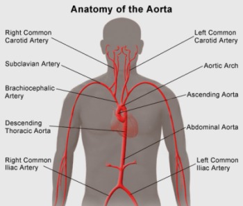
Figure 1: The aorta with its different main branches
The
aorta is the largest blood vessel in the human body and serves as the main artery directing oxygenated blood from the heart to the rest of the body. The aorta arises from the left side of the heart (the left ventricle), arches and moves down through the length of the body till the lower part of the abdomen, where it then divides into two major branches that run down each lower limb.
As it is such a large continuous vessel, the aorta is divided into different sections to easily pinpoint where along the vessel is being referred. These sections are the:
- Ascending aorta
- Aortic arch
- Descending aorta
- Abdominal aorta
The aorta also gives off smaller branches along its path through the body. These branches break off into smaller branches until they reach the specific organs, tissues and cells to deliver the oxygenated blood. The simplest way to describe the aorta is by imagining a highway that has multiple exits that give into smaller roads and side roads.
The aorta and all its branches are called arteries. Arteries have elastic walls that allow them to respond to the pressure caused by the blood ejecting from the left side of the heart. The elastic wall itself has three layers.
What is aortic aneurysm?
An
aneurysm happens when a section of an artery experiences weakness in its arterial walls. This causes that part of the artery to swell out like a balloon.
An aneurysm can occur congenitally (when someone is born with an aneurysm) or as a result of a disease or an injury to the artery.
Therefore, an
aortic aneurysm is when a part of the aorta's wall is weak, causing it to stretch and balloon out. Though an aortic aneurysm may occasionally cause some discomfort, the main concern is that as the wall stretches more, it becomes thinner and poses a risk of rupture. A ruptured aneurysm can be life threatening as it can cause massive internal bleeding.
What is an aortic dissection?
An
aortic dissection refers to a tear in the elastic wall of the aorta.
The exact cause of an aortic dissection is not known but some of its risk factors are:
- Ageing
- Atherosclerosis
- Blunt trauma in the chest area
- High blood pressure
The tear when it occurs is often due to the damage of the inner lining of the aorta and is more common along the part of the aorta that travels in the chest area. When the tear happens in the aortic wall, the layers within the wall are forced apart by the blood entering into these tears. If the tears completely dissect (cut through) the aorta, massive and rapid blood loss occurs. Therefore, an aortic dissection is considered a medical emergency.
The diagnosis of an aortic aneurysm or aortic dissection is similar. There are different methods available to not only diagnose these conditions but to also help doctors to determine the best treatment options and follow-up plans.
1. Chest X-Ray (CXR)
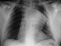
CXRs may help to diagnose thoracic aneurysm or dissection.
Although they are not the best available method to diagnose these conditions, it is easily available and can help suggest and alert doctors of an abnormality of the aorta and prompt further tests.
1.1. Abdominal X-ray
Abdominal X-ray can sometimes diagnose aortic aneurysm but it is not very accurate.
2. CT Scan (Angiograms of aorta)

Computed Tomography scans (CT scans), also known as "CAT Scan", is the preferred method of diagnosing an aortic abnormality through imaging.
However, to ensure a better image, a contrast medium (dye) is given intravenously before the image is taken. Therefore, this method may not be used in some patients, such as, those with known allergies or kidney problems, for example.
This is now the standard for preparing and planning patients for surgery (both endovascular and open).
3. Echocardiograms
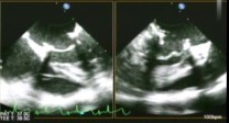
An echocardiogram also known simply as an "ECHO", is basically an ultrasound for your heart that is able to take images of the heart as it pumps.
An echo helps to give a clearer picture of what is happening to the heart and the aorta and is useful especially when evaluating certain parts of the heart and the aorta.
It is also part of the work up to check the heart function before a surgery.
4. Magnetic Resonance Imaging (MRI)
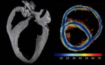
Magnetic resonance imaging (MRI) is similar to a CT scan. However, one of its benefits is that it does not use radiation to capture images. Instead it uses a powerful magnetic field to do so. Another benefit is that the contrast medium (dye) used in an MRI seems to be better tolerated in patients.
5. Aortic Angiography
Aortic angiography is a procedure, which uses a contrast medium (dye) that is injected into the aorta and x-rays that are taken to determine how blood is flowing inside it.
Symptoms of an aortic aneurysm
If aneurysm develops slowly over many years, they are often asymptomatic (no symptoms).
However, if the aneurysm occurs in an artery that is near the surface of your skin, you will notice a growing bulge or mass that seems to throb. These can be painful.
When an aneurysm grows quickly or if it ruptures, the symptoms can occur suddenly. There will be pain and symptoms of shock due to blood loss such as:
- Clammy skin
- Dizziness
- Nausea and vomiting
- Rapid heart rate
- Low blood pressure
Symptoms of an aortic dissection
Most of the time, the symptoms of an aortic dissection happen suddenly. These include:
- Severe chest pain that can feel like a heart attack.
- Pain that has been described as a sharp, stabbing, tearing or ripping pain.
- It can be felt below the chest bone (sternum) and moves under the shoulder blades or to the back.
- The pain may also move to the shoulder, neck, arm, jaw, abdomen or the hips.
- It has been described as a "moving pain", starting from the arms to the legs as the dissection worsens.
Symptoms of shock due to massive blood loss such as:
- Dizziness
- Clammy skin
- Nausea and vomiting
- Pallor - when the skin becomes very pale
- Rapid and weak pulse
- Shortness of breath and difficulty breathing when lying down
Treatment for aortic aneurysm
Aortic aneurysm treatment depends on the size, location and the patient's state of health.
For example, if your aneurysm is small and there are no symptoms, your doctor will probably recommend a "watch and wait" approach. Here, regular scheduled appointments are made where any changes in the aneurysm is monitored by the various imaging methods such as CT scan or MRI.
However, if the aortic aneurysm is large or grows more than 1 cm per year, then surgery may be recommended for you.
1. Aortic Surgery
There are various types of aortic surgery that are available to treat aortic aneurysm.
1.1. Open Abdominal or Open Chest Surgery
This is the standard surgery when the aortic aneurysm reaches the need for surgery. The procedure involves the replacement of the portion of the aorta that is swollen, with an artificial graft. The graft is made from a material that does not wear out and is sewn in place with a permanent suture material.
1.2. Endovascular Surgery
Compared to open surgery explained above, endovascular surgery allows for the repair of an aortic aneurysm with less trauma to the aorta, less blood loss and fewer days in intensive care during recovery.
Although it usually benefits those who are at high risk of complications during surgery, it may not be suitable for every person. Your doctor will be able to decide if this form of surgery is the better option for you.
During the procedure, a thin tube (or catheter) is inserted into the blood vessel via an artery in the leg. Attached to the catheter is a graft that is placed where the aneurysm is located using x-ray guidance. Once in place, the graft is expanded. The metal frame of the graft expands like a spring which serves to hold up the walls of the aorta. This graft reinforces the area of the weakened artery wall and prevents the risk of subsequent rupture of the aneurysm. The blood flow is directed away from the aneurysm and flows through the graft instead.
1.3. Hybrid Elephant Trunk Procedure
This procedure is done in patients with large aortic aneurysms located in various sections of the aorta. It is a more complex procedure that involves two stages.
2. How should you prepare for the surgery?
- Let your doctor know beforehand what medications you are on as some of these medications may have to be stopped before the surgery.
- Smokers need to stop smoking before the procedure.
- Give your doctor a list of your medical history, especially if you have any allergies.
- You will be informed if you would need to undergo other tests prior to surgery, some tests you will need are:
- Electrocardiogram (ECG)
- Blood tests
- Chest X-ray
- Urine sample
- Your doctor will explain the details of the procedure to you and answer your questions should you have any.
2.1. What happens before the surgery?
- Do not eat or drink for 6-8 hours before the surgery.
- You will be asked to sign a consent form.
- On the morning of the surgery, you will be asked to remove all jewelry, dentures and undergarments.
- Empty your bladder before the preoperative medication is given, which is to help you relax.
2.2. What happens during the surgery?
- You will be given anaesthesia after entering the operating theatre.
- An incision will be made down the middle of the chest.
- Your breastbone will be separated to allow the surgeon to examine your heart.
- Your heart will be connected to the heart-lung bypass machine. This machine supplies oxygen to your blood and pumps it back to the rest of your body.
- Now, the aortic surgery begins.
- The aorta is gently opened to reveal your aortic valve.
- The replacement valve will be sewn in place after the old valve is removed.
- After the aorta has been closed with stitches, you will slowly be removed from the heart-lung bypass machine after your heart regains strength.
2.3. What are the potential risks of this surgery?
Minor complications include:
- Nausea and vomiting
- Infection at the operated site
- Minor bleeding
- Allergic reactions to the plaster or certain medications
Major complications include:
- More severe infection
- Bleeding during or after the surgery
- Heart attack
- Stroke
Though no surgery or procedure is risk free, aortic surgeries have been performed for many years with good results and limited complications. Your doctor, prior to you giving consent for the surgery, will explain these and other risks in detail.
2.4. What happens after the surgery?
- Immediately after the surgery, you will be sent to the intensive care unit (ICU), where you will be taken care of for several days or as long as it is needed, until you are stable enough to be transferred to the ward.
- In an ICU, your heart rate, blood pressure and breathing are monitored continuously.
- Physical therapy will start as soon as possible to allow for a faster recovery. For example, you will learn how to move your upper arms without hurting your breastbone while it heals.
- You may also receive counselling on how to live a healthier lifestyle.
2.5. Going home
- Have a relative or friend pick you up on the day of your discharge from the hospital.
- If you live alone, you may want to think about having someone to help you with some of your daily activities until you have completely healed.
- Ensure that you remember your follow-up appointments with your doctor and make sure that you take any medications given to you on discharge.
3. Aortic Dissection treatment
The treatment for aortic dissection depends on where in the aorta in occurs. Regardless, it is a life-threatening condition that has to be treated as soon as possible.
- If the dissection occurs in the area of the aorta that arises from the heart (the ascending aorta) then treatment is surgery.
- However, if the dissection occurs in other parts of (the descending/abdominal) aorta, it can be managed with either medications or surgery.
- Of the people with a ruptured aorta, less than half of them survive.
3.1. Types of surgery
3.2. Medications
- Beta-blockers — this is a category of drugs used to lower the blood pressure. They are given intravenously and are the first drugs of choice.
- Pain relief — strong pain medications are also given to make the patient comfortable when in severe pain.
3.3. What happens after treatment?
- Once the dissection heals either through surgery or medications, your blood pressure will have to be treated well.
- There will also be life-long monitoring of the area where the dissection occurred. This is because there is a risk that a previously dissected descending aorta may enlarge and rupture.